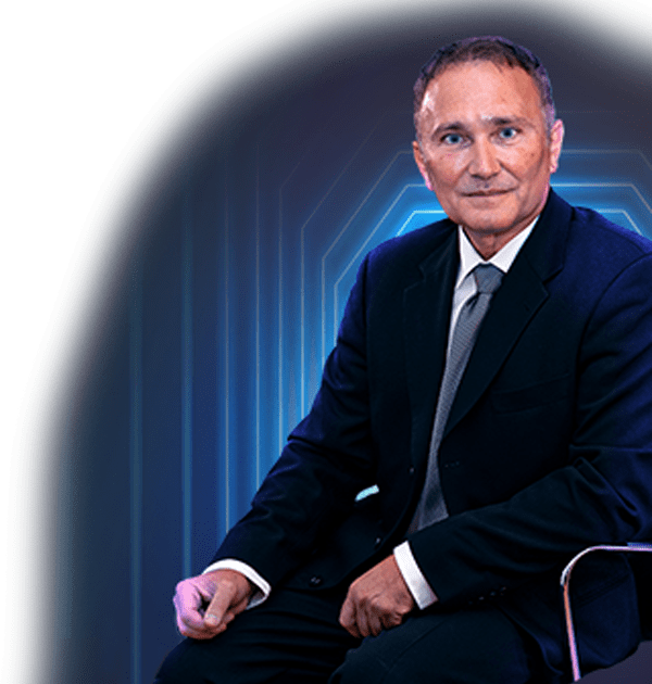
Welcome!

EMG Laboratory, led by Dr. Drasko Simovic, is a premier medical practice with an exclusive focus on diagnostics of nerve and muscle disorders. We have been serving the medical communities in Massachusetts and Southern New Hampshire for decades by providing the highest levels of patient care and diagnostic support for referring professionals.

Practice exclusively
dedicated to
EMGs since 1997

Board
Certified in
four disciplines

Highest level of
National EMG Lab
Accreditation

Information for patients
about the EMG Test

Do you have pain, numbness, tingling, burning or weakness in your limbs?

If yes, ask your doctor if our EMG Lab can help diagnose your condition!

Why do referring offices choose us?

Impeccable
service to the
refering offices

Urgent cases
are rapidly
accommodated

All EMG Tests
are performed by
Dr. Simovic

Quality and
reliability of the
EMG Test results

All unexpected
cases are personally
discussed

EMG Test results
are promptly
reported

When can an EMG help?

Electrodiagnostic medicine helps diagnose conditions with symptoms including pain, nubmness, burning, tingling and / or weakness. The primary goal of electrodiagnostic examination is to determine the site of the lesion. Such testing serves a variety of functions:

•  To establish the diagnosis
•  To evaluate for concomitant conditions
•  To argue for or against an alternative diagnosis
•  To establish the type and quantify the severity of the condition

By establishing the correct diagnosis we can help select the best therapeutic option. Although clinical evaluation is irreplaceable, electromyography can help in providing in-depth information about the state of the peripheral myelin, axons or muscles.

Leave us a review

EMG and NCS

Electromyography (EMG) and nerve conduction studies (NCS) are tests that measure the electrical activity of muscles and nerves. Nerves send out electrical signals to make your muscles react in certain ways. Nerves also send signals from your skin to your brain, which then processes the signals, so you can experience a variety of skin sensations.

  • An EMG Test looks at the electrical signals your muscles make when they are at rest and when they are being used.
  • A Nerve Conduction Study measures how fast and how well the body’s electrical signals travel along your nerves. EMG and nerve conduction studies are used to help diagnose a variety of muscle and nerve disorders and to quantify the severity of your condition.


An EMG test helps find out if muscles are responding the right way to nerve signals. Nerve conduction studies help diagnose nerve damage or disease. When EMG tests and nerve conduction studies are done together, they help doctors tell if your symptoms are caused by a muscle disorder or a nerve problem. With EMG and NCS studies, your doctor can next select the best therapeutic option to help you.

Medical illustrations on this website are attributed to: Freepik.com and author Kjpargeter

Lateral Femoral Cutaneous Nerve

From: www.wheelessonline.com 

– Anatomy:    

– LFCN, as its name suggests, is purely sensory;    
– it arises from L2 and L3, travels downward lateral to the psoas muscle, crosses the iliacus muscle (deep to fascia), passes either thru or underneath the lateral aspect of the inguinal ligament, and finally travels onto innervate the lateral thigh;
– it divides into anterior and posterior branches and supplies skin on lateral aspect of thigh;
– in the study by Hospodar et al (JTO 1999), the course of the nerve was variable, but was most commonly found at 10-15 mm from the ASIS and as far medially as 46 mm from the ASIS;
– in no specimen did the nerve pass lateral to the ASIS (eventhough historically the nerve is thought to pass lateral to the ASIS in 10% of population);
– in all specimens the nerve passed underneath the ilioginal ligament and anterior to the iliacus muscle; 

– Meralgia Paresthetica: 

– entrapment syndrome of the lateral femoral cutaneous nerve causing burning, numbness, and paresthesias down the proximal-lateral aspect of the thigh;
– may be idiopathic, be a result of trauma, previous operations, and in some cases may arise from Perthes Disease abduction splints;
– in idiopathic cases, the nerve may be encased in bone by the growing apophysis of the anterior superior iliac spine, or may be entrapped in fascia either proximal or distal to the ASIS;    
– diagnosis is made by:           
– reproduction of the pain by deep palpation just below the anterior superior iliac spine and by hip extension;           
– relief of pain by localized injection of lidocaine;    
– treatment: when diagnosis is not in doubt and the symptoms are severe, consider operative decompression at the site of constriction;